- Our Breakthrough Technology: GlucoBeam
- GlucoBeam Key Features
- More about RSP’s Raman system
- Raman Systems Schematic
- GlucoBeam in Action
- Intellectual Property
- More about RSP's Critical Depth Raman
- Publications
-
Our Breakthrough Technology: GlucoBeam
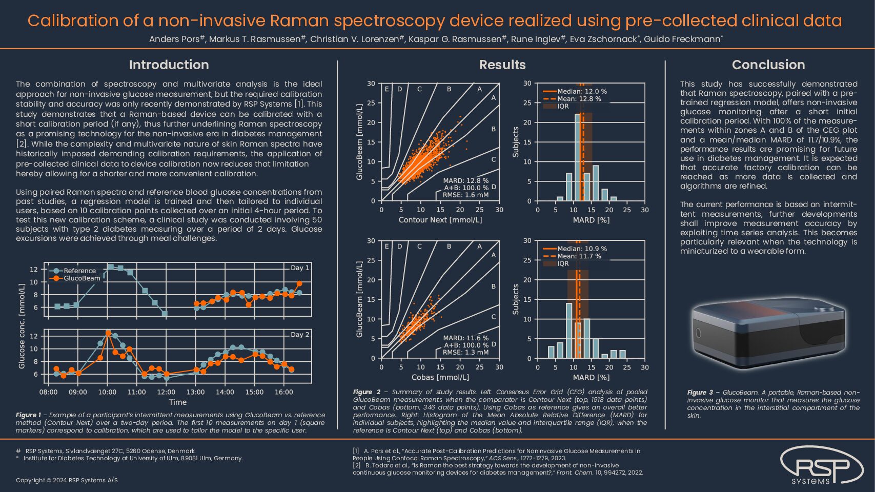
Over the past 15 years, RSP has developed a clinically proven device that measures blood glucose in an entirely pain-free manner by using a well-understood principle in a new way. This breakthrough non-invasive glucose monitor will address the unmet need for diabetes self-management
The innovation of our device comes from combining Raman Spectroscopy with Critical depth technology.
-
GlucoBeam Key Features
A non-invasive glucose monitor that uses a beam of light to painlessly and accurately measure glucose in the interstitial fluid. GlucoBeam has the following key features:
- Reputable Technology
Fine-Tuned over 10 Years of Testing. - Advanced Raman spectroscopic technology, optimised for glucose measurement
- Overcome numerous technological barriers
- Reliable Performance
Proven in Unsupervised Home Use
- Tested in >550 patients in clinical and home settings
- Strong correspondence between GlucoBeam measurements and reference blood glucose measurements
- Real-Life Utility
Developed with Thorough Consideration of User Experience
- Simple and easy-to-use design
The illustration above visualizes the penetration of near-infrared light in the skin and how photons may undergo scattering events, thus eventually (if not absorbed) leaving the skin with lateral displacement and at large angles. Furthermore, it is important to note that the dynamic glucose signal resides in the living part of the skin (living epidermis and dermis) and, for this reason, the Raman instrumentation should exclude the signal from the top skin layer (stratum corneum). In practice, this is achieved using the confocal principle that allows for depth selectivity. Last but not least, it should be emphasized that a biological Raman spectrum features a multitude of Raman lines from many different molecules and, consequently, it requires advanced multivariate analysis techniques to quantify the concentration of a specific molecule.
- Reputable Technology
-
More about RSP’s Raman system
What is Raman Spectroscopy?
It is a well-known method of molecular identification and quantification, based on the Nobel Prize-winning Raman scattering principle. A Raman spectrum, measured from a sample, acts like a “structural fingerprint”. Raman spectroscopy has increasing applications in health care and diagnostics.
RSP: the first company to overcome Raman spectroscopy challenges
- Widespread use of this optical technique was deterred due to its weak optical process.1 Only one in a million incident photons experience the Raman scattering
- RSP has overcome critical problems such as a low number of photons and strong background signals by introducing a confocal optical system. This system together with lens objectives collects photons over a large solid angle
- We are also proud of our spectrometer, which features high throughput and resolving power to various Raman lines
- This optical system features a laser wavelength of 700-850 nm to maximize the number of photons generated
- It is also important to note that the dynamic glucose signal resides in the living part of the skin (living epidermis and dermis) and, therefore, the Raman instrumentation should exclude the signal from the top skin layer (stratum corneum).1 In practice, this is achieved using the confocal principle that allows for depth selectivity.
- Lastly, a biological Raman spectrum features a multitude of Raman lines from many different molecules and, consequently, it requires advanced multivariate analysis techniques to quantify the concentration of a specific molecule.
1. Lundsgaard-Nielsen SM et al. PloS one. 2018; 13: e0197134
-
Raman Systems Schematic
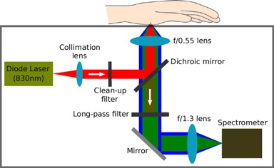
A Raman system consists of three central hardware components: an excitation laser, a light guiding probe optics unit and a spectral dispersing element with a spectrometer to detect light.
It has a frequency-stabilized, solid state laser, which is small, inexpensive and rugged. The spectrometer analyzes the Raman scattered light from the sample being transmitted via the probe optics.
Every component is optimized and standardized for home use.
-
GlucoBeam in Action
This innovative self-management device is extremely easy
1) User contacts device with base of thumb (the thenar) and laser is emitted into skin2) Raman scatter from glucose molecules in interstitial fluid is collected and analysed
3) Glucose concentration will be displayed in a fraction of a minute
In the plot in black below, a Raman spectrum of Thenar skin background subtracted and average over 121 spectra with an exposure time of 10 sec is seen. In red a scaled Raman spectrum of glucose/water solution 555.5 mmol/l with an exposure time 13 sec. Both acquired with the WM34 data-mining equipment.
-
Intellectual Property
Through our highly skilled team, we have advanced the technological application of Raman spectroscopy to be used to non-invasively measure relevant molecules in human tissue.
These advancements are a result of nine years of R&D and pre-clinical studies where we refined Raman technology, focusing on improving the optical system, electronics and data analysis algorithms.
Furthermore, we have carefully developed the know-how of in-vivo measurements required to:
- Selectively and efficiently collect Raman signal from the interstitial fluid and cells
- Analyze spectral data to derive the concentration of a given molecule
- Build a calibration model and use it for deriving concentration values from measurements
In the process, we have discovered and patented fundamental aspects of Raman detection of glucose and other substances in human tissue. This removes the dependence of signal calibration on the probe position, allowing for unprecedented performance.
-
More about RSP's Critical Depth Raman
RSP’s critical depth technology is a proprietary method to measure glucose at a specific depth within the skin (the interstitial compartment)1
This technology shows strong analytical performance and stable calibration
RSP Systems utilizes a confocal optical design enabling Raman spectroscopy measurements from skin layers below the top layer (Stratum Corneum). By excluding the optical signal from the upper skin layers and lower layers (Reticular Dermis).
A confocal optical system using a spatial pinhole to block light originating from out-of-focus regions in the sample. The lenses are designed in such a way that the excitation light is focused on the desired depth and light collected from the specific depth is guided through the pinhole. Preferable it is only light originating from the focal plane that is collected.
1. Lundsgaard-Nielsen SM et al. PloS one. 2018; 13: e0197134
-
Publications
- Poster ATTD 2020
Non-invasive glucose monitoring by a raman spectroscopybased prototype after meal challenge in Type 1 diabetes - Poster DTM 2019
Measurement accuracy of a newly developed prototype system for non-invasive glucose monitoring - Abstract ADA 2019
Measurement accuracy of a newly developed prototype system for noninvasive glucose monitoring - Poster ADA 2019
Measurement accuracy of a newly developed prototype system for non-invasive glucose monitoring - Poster DTM 2018
Non-invasive Raman-based Glucose Monitoring with weeks of sustained calibration - Lundsgaard-Nielsen SM et al. PloS one. 2018; 13: e0197134
- Poster ATTD 2020
A tactile design concept with risk mitigation features

Features